 Site Search


Cytopathology Case 13
FNA of a mesenteric mass in a 53 year-old male
by Leslie E Lopez, MD
Editor: Benjamin L. Witt, MD, Clinical Instructor of Pathology, University of Utah, and Medical Director, Cytopathology, ARUP Laboratories
A 53 year-old male with multiple medical problems including type 2 diabetes mellitus, coronary artery disease status post myocardial infarction, and epilepsy was transferred to our care for evaluation of a progressive right thigh mass and the recent complaint of gastrointestinal discomfort.
Surveillance abdominal imaging demonstrates two mesenteric-based masses measuring 8.6 cm and 4.0 cm in greatest dimension. In addition, a deeply seated, 14.0 cm right thigh mass is evident on an MRI of the leg.
After clinical evaluation, resection of the thigh mass was scheduled, as well as a fine needle aspiration (FNA) of the larger mesenteric-based mass to discern whether the two represented the same process or were unrelated.
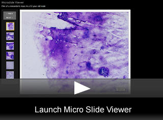
The cytology images are from the FNA of the mesenteric mass. The histology images are from the right thigh mass.
Cytomorphology Description:
Microscopic Features:
- Low power smear shows abundant magenta colored matrix associated with cell clusters(fig. 1)
- A higher power view of the cellular areas demonstrating a proliferation of round to oval cells with prominent fat differentiation and an associated vascular network(fig. 2).
- A view showing the proliferating cells, fatty differentiation, with associated vessels and matrix(fig. 3).
- A different area of the smear showing similar features(fig. 4).
- On histology the low cellularity proliferation of oval to round cells having adipoctyic differentiation, an associated capillary network, and abundant light pink myxoid matrix match the features noted on cytology(fig. 5).
- High power view shows multiple candidate lipoblasts with two in the lower left field of view(fig. 6).
Final Diagnosis & Discussion
© The copyright for photographs and digital images shown in this case report is owned jointly by Benjamin L. Witt, MD and ARUP Laboratories, Inc. Unlicensed publication in print, on the internet, or in any other media form of these digital images or photomicrographs for any purpose without written permission is strictly prohibited. Limited use for teaching is permitted. Please contact Kyle Harris for licenses and permissions. If you wish to use these images as aid in lectures or scientific slide presentations, each image should accompany the following text: "copyrighted material: www.arup.utah.edu"