

 Connect Sign In
EUS FNA of a Gastric Mass in a 61-Year-Old Male
Magda Esebua, MD, Assistant Professor of Pathology, University of Missouri-Columbia
Editor: Brian T. Collins, MD, Professor of Pathology, University of Utah, and Medical Director, Cytopathology, ARUP Laboratories
A 61-year-old, white male presented with abdominal pain and early satiety. There was no chest pain, reflux, or shortness of breath. Physical exam revealed no palpable abdominal masses.
As part of the workup, esophagogastroduodenoscopy was performed, revealing a gastric sub-mucosal mass at the antrum measuring 3.5 cm. This mass was arising from muscularis propria and had two lobules. Endoscopic ultrasound-guided fine needle aspiration (EUS FNA) was performed. Six alcohol fixed slides and 30 cc of cytolyte were sent to cytology lab for proccessing and examination.
EUS FNA cytology of the gastric mass showed a large cell neoplasm most consistent with gastrointestinal stromal tumor. Approximately two months later a partial gastrectomy was performed and the specimen was sent for histologic evaluation.
Micro Slide Viewer
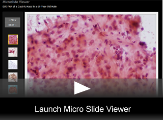
Cytologic Features:
- The cytology laboratory received 6 alcohol fixed smears and 30 cc of cytolyte. Papanicolaou stained smears; one cell block and one liquid-based slide were prepared. Papanicolaou stained smears showed a cellular sample composed of loosely cohesive groups of neoplastic cells. These neoplastic cells were predominantly epitheliod with eccentric oval to round nuclei and moderate to abundant eosinophilic or amphophilic granular cytoplasm with ill defined borders (fig. 1).
- Cells were imbedded in myxoid stroma and rare transgressing capillaries were noted. Prominent intranuclear inclusions and binucleation were present (fig. 2).
- The cell block preparation was moderately cellular; cells were arranged in sheets and showed cytologic features similar to what was present on the slides.
- An immunocytochemical panel using serveral antibodies was performed: CD117(c-kit), CD-34, smooth muscle actin (SMA), S-100, MART-1, CK-7 and CK-20, LCA.(fig. 3).
- Neoplastic cells were negative for CD117, S-100, A-103, CK-7, CK-20 and LCA. Neoplastic cells were positive for CD-34 (fig. 4).
Histologic Features:
- Partial gastrectomy specimen received in histology lab showed an endophytic submucosal neoplasm. (fig. 5).
- Sectioning revealed 3.5 by 3.2 by 3.0 cm multi-lobulated well circumscribed tan-gray tumor confined to the wall of the stomach.
- Hematoxylin & Eosin stained slides show a neoplasm composed of predominantly large epitheliod cells with pleomorphic nuclei, prominent small nucleioli and abundant eosinophilic to clear cytoplasm.(fig. 6).
- Binucleation as well as multi-nucleation were noted. Occasional cytoplasmic vacuoles and intranuclear inclusions were also present. One mitosis was identified per 50 400X high power fields. No necrosis was seen.
- Immunohistochemical stains were as follows; CD117 negative (fig. 7) CK, Vimentin, EMA, CD-31, Mart-1, HMB-45 and S-100 negative; CD-34 positive,(fig. 8), and SMA weakly positive.
DISCUSSION
Gastrointestinal stromal tumors (GIST) are the most common mesenchymal tumors of the gastrointestinal tract. The majority of these tumors are found in the stomach. The cell of origin is considered to be the intestinal pacemaker cell, the cell of Cajal. GISTs are usually accompanied by a somatic mutation of CD-117(c-kit), which is a tyrosine kinase receptor normally expressed by the intestinal cells of Cajal. The specific diagnosis of GIST is important because of the possible response to tyrosine kinase inhibitor, imatinib mesylate (Gleevec).
GISTs show two basic cells types: spindle and epithelioid. Epithelioid variants occur in approximately 25-30% of gastric GISTs and correspond to tumors previously termed “leiomyoblastomas” or “epithelioid leiomyosarcoma”.
The cytology depends upon the cell type, but there are no pathognomonic cytologic features of GIST. In epitheliod GISTs, cytologic smears are usually cellular, and the cells appear predominantly singly or in loose clusters. Upon processing with Papanicolaou stain, the epitheliod cells contain moderate amount of amphophilic to eosinophilic cytoplasm. Nuclei are round or oval and vesicular. Intranuclear inclusions and binucleation are frequent findings. Small nucleoli may also be present. Stroma is usually myxoid and transgressing capillaries may be seen.
The most important immunocytochemical marker of GIST is expression of c-kit (CD117), which is found in vast majority (>90 percent) of GISTs. Staining is usually strong and widespread. It may show a variety of patterns including diffuse cytoplasmic staining, localized perinuclear dote and membranous.
Most (>80 percent) gastric GISTs also express CD34, but smooth muscle actin is demonstrable in about 25 percent.
Some GISTs can be “KIT-negative”, despite having typical morphological (and other immunohistochemical) features.
This is an unusual case of epithelioid variant of GIST which was negative for c-kit (CD117) on both FNA and surgical resection. Significant pitfalls for the diagnosis of epitheliod GIST include neuroendocrine tumors, carcinomas and melanomas, and other neoplasms with an epitheliod appearance. Differential diagnosis with neuroendocrine tumors may be challenging because of the plasmacytoid appearance of cells along with round nuclei containing finely granular chromatin. Immunocytochemical analysis (with chromogranin or synaptophisin) can also be helpful in determining the diagnosis.
When the GIST is composed of discohesive tumor cells with eccentric large nuclei, prominent nucleoli, and intranuclear inclusions, melanoma becomes an important differential diagnosis. However GISTs are usually negative for the common melanoma markers such as S-100, Mart-1, and HMB-45.
While it is prudent for a cytopathologist to consider GIST in the differential diagnosis of any unusual gastric neoplasm with epithelioid features, it is important to realize that not all GISTs are c-kit (CD-117) positive.
References
- WHO Classification of Tumours of Digestive System: IARC Press, Lyon, 2000.
- DeMay M. Practical Principles of Cytopathology. 2007.
- Dong Q, McKee G, Pitman M, Geisinger K, Tambauret R. Epithelioid variant of gastrointestinal stromal tumor: Diagnosis by fine needle aspiration. Diagnostic cytopathology. 29(2):55-60.
- Lograno R, et al. Imaging, morphologic, and immunohistochemical correlation in gastrointestinal stromal tumors. Cancer Cytopathology. 2006;June:257-267.
© The copyright for photographs and digital images shown in this case report is owned jointly by Brian Collins, MD and ARUP Laboratories, Inc. Unlicensed publication in print, on the internet, or in any other media form of these digital images or photomicrographs for any purpose without written permission is strictly prohibited. Limited use for teaching is permitted. Please contact The Webmaster for licenses and permissions. If you wish to use these images as aid in lectures or scientific slide presentations, each image should accompany the following text: "copyrighted material: www.arup.utah.edu"
