 Site Search


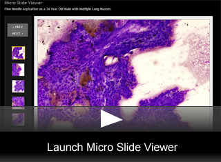
Fine Needle Aspiration on a 34-Year-Old Male with Multiple Lung Masses
by Julie Ann Walby, MD, Cytopathology Fellow, University of Utah
Editor: Brian T. Collins, MD, Professor of Pathology, University of Utah, and Medical Director, Cytopathology, ARUP Laboratories
A 34-year-old previously healthy male presents with multiple bilateral lung nodules. An outside spiral CT report identifies a prominent medial right hemi-thorax mass, measuring 12.8 cm in greatest dimension which is compressing the right atrium. Additional lesions are found in the lateral right lung apex measuring in greatest dimension 3.8 cm, left upper lobe measuring 3.3 cm, and lateral right lower lobe measuring 2.6 cm. There is no consolidation, pleural effusion or pneumothorax present. There are multiple prominent anterior-superior mediastinal lymph nodes but all are normal by size criteria. There are no bone lesions or any lesions below the diaphragm on an abdominal CT. A CT-guided fine needle aspiration was performed on the right lung mass. One pass was done with a single smear for modified Wright-Giemsa staining and a cell block prepared. Based on the patient history and the morphologic features of the cells, immunohistochemical stains were performed on the cell block.
FNA Findings
The modified Wright-Giemsa smears consist of large groups of cells in a background of hemorrhage and necrosis. (fig. 1) The groups are loosely cohesive and are composed of a two cell population. (fig. 2) The cells located centrally are moderate in size, round to polygonal in shape. Cell borders are somewhat distinct. Nuclei of these cells are single and bland with mild pleomorphism. The nuclei are centrally located in a pale to clear cytoplasm. (fig. 3) Nucleoli are indistinct in air dried smears. The second population of cell is peripherally located. These are giant cells that are multinucleated with nuclei that are large and atypical. (fig. 4) The cytoplasm is abundant and eosinophilic, vacuolations are occasionally present. The chromatin is smudged but prominent nucleoli can be seen. (fig. 5) The cell block highlights the multinucleated giant cells with eosinophilic cytoplasm. (fig. 6) Immunohistochemical stain is shown. (fig. 7)
Final Diagnosis & Discussion
© The copyright for photographs and digital images shown in this case report is owned jointly by Brian Collins, MD and ARUP Laboratories, Inc. Unlicensed publication in print, on the internet, or in any other media form of these digital images or photomicrographs for any purpose without written permission is strictly prohibited. Limited use for teaching is permitted. Please contact The Webmaster for licenses and permissions. If you wish to use these images as aid in lectures or scientific slide presentations, each image should accompany the following text: "copyrighted material: www.arup.utah.edu"