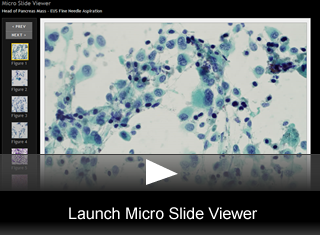
Head of Pancreas Mass—EUS Fine-Needle Aspiration
by Julie Ann Walby, MD, Cytopathology Fellow, University of Utah
Editor: Brian T. Collins, MD, Professor of Pathology and Medical Director, Cytopathology, ARUP Laboraotires
A 57-year-old male presents to the emergency room with a 1-month history of flu-like symptoms, including epigastric pain that has been increasing and is now constant for the past week. This pain is dull and somewhat boring with some radiation to the back. Jaundice has recently been noticed and is reportedly increasing. He has also experienced a 10 lb weight loss over the past 6 months. Stools are reportedly acholic and fatty, while his urine has been darkening over the past month. He has no significant past medical history. He does not smoke and currently consumes 2 to 3 beers daily. Abnormal laboratory results include profoundly elevated hepatic transaminases and a bilirubin of 12.8, CEA of 3.1, and CA19-9 of 120. Ultrasound reveals a mass in the head of the pancreas with dilated biliary ducts. A CT scan shows an ill-defined, hypodense, hypermetabolic, solid mass in the head of the pancreas abutting the inferior margin of the portal vein, measuring 2.8 cm in greatest dimension. Also noted is possible gall bladder involvement. There is no evidence of metastatic disease. An endoscopic ultrasound-guided fine-needle aspiration was performed. At the time of this procedure, the mass was noted to measure 3.7 cm and to be somewhat bi-lobed.
Micro Slide Viewer
FNA Findings
The smears are cellular with discohesive cells which are clearly recognized as malignant (fig. 1). Some cells are multinucleated giant cells but not of the osteoclastic type (fig. 2). The cells are large and pleomorphic with bizarre forms. There are occasional isolated, large, atypical columnar “tombstone cells” identified (fig. 3). The nuclei are hyperchromatic and are often eccentric with prominent nucleoli (fig. 4). The cytoplasm of other cells is stripped or transparent (fig. 5). There is an inflammatory infiltrate present with emperipolesis. Anisokaryotic daughter nuclei and cellular cannibalism can be seen. Some spindle-shaped cells are found, but there is no recognizable pigment present. The cytoplasm of many cells is dense and eosinophilic, with frequent mitoses (fig. 6). A background of necrosis, hemorrhage, and cystic degeneration is common.
 Site Search


