Cytopathology Case 16
FNA and Core Biopsy Diagnosis of a Retroperitoneal Mass
by Irene Czyszczon, DO
Editor: Benjamin L. Witt, MD, Medical Director, Cytopathology, ARUP Laboratories, and Assistant Professor of Pathology, University of Utah.
A 65-year-old female presented to her physician with abdominal swelling and episodic abdominal pain for 1 year duration. Past medical history includes hypertension and hysterectomy for unknown reason. Physical exam shows abdominal fullness and leg swelling. A CT scan reveals a 16 cm retroperitoneal mass which displaces the kidney and compresses the adrenal gland. The radiologist on site indicated that the mass seems to be closely associated with the vena cava. The patient underwent ultrasound-guided FNA and core biopsy.
Cytomorphology Description:
Microscopic Features:
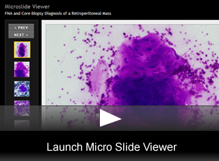
- Aspiration cytology shows moderately cellular, tight clusters of spindle cells with a subtle metachromatic stroma. The nuclei are elongated. While a few have pointed ends, the majority are plump with blunted ends and show occasional indentation. Nucleoli are in general inconspicuous but occasionally distinct.(fig. 1), (fig. 2), (fig. 3), (fig. 4)
- Histology shows a hypercellular specimen featuring spindle cells arranged in sweeping, intersecting fascicles. Cytoplasm is densely eosinophilic and shows frequent vacuolization. Nuclear atypia is present and is demonstrated by hyperchromasia, irregularity and enlargement.(fig. 5), (fig. 6)
- Immunohistochemical staining for SMA shows diffuse positivity in the tumor cells.(fig. 7)
- Further immunohistochemical stains show the tumor cells to be positive for desmin, while they are negative for S100.
Final Diagnosis & Discussion
© The copyright for photographs and digital images shown in this case report is owned jointly by Benjamin L. Witt, MD and ARUP Laboratories, Inc. Unlicensed publication in print, on the internet, or in any other media form of these digital images or photomicrographs for any purpose without written permission is strictly prohibited. Limited use for teaching is permitted. Please contact The Webmaster for licenses and permissions. If you wish to use these images as aid in lectures or scientific slide presentations, each image should accompany the following text: "copyrighted material: www.arup.utah.edu"
 Site Search


