

 Connect Sign In
Fine Needle Aspiration of 81-Year-Old Female With an Adrenal Mass
Brent J. Huddleston, MD Fellow, Cytopathology. University of Utah, Department of Pathology
Editor: Brian T. Collins, MD, Professor of Pathology, University of Utah, and Medical Director, Cytopathology, ARUP Laboratories
An 81-year-old female residing in a skilled nursing facility has a past medical history significant for hypertension, diabetes mellitus, hypothyroidism, hyperlipidemia, and coronary artery disease. The patient complains of an approximately 5-month history of weakness, fatigue, decreased oral intake, 20-pound weight loss, night sweats, nausea, vomiting, and occasional syncope. These have all been getting worse over time. The syncope occurs while standing or sitting and she has had approximately 3 to 4 episodes of complete blackout. She also complains of occasional chest pain with lightheadedness and palpitations. Her orthopedic surgeon saw her for back pain and felt a lump in her axilla and neck, and ordered an MRI of her abdomen that revealed a right adrenal mass. CT-guided fine needle aspiration was performed of the adrenal mass.
Micro Slide Viewer
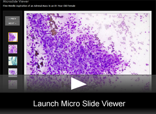
Fine needle aspiration shows the following:
- The smears are cellular with mostly cohesive groups of cells (fig. 1).
- Some discohesive and loose acinar patterns are seen (fig. 2).
- There is a prominent vascular network of capillaries within the larger cell groups (fig. 3).
- The nuclei are mostly round to oval (fig. 4) with some scattered pseudonuclear inclusions (fig. 5).
- There is moderate anisonucleosis with some binucleate forms present. Nucleoli are prominent (fig. 3).
- The cytoplasm is abundant, granular, with indistinct cell borders, and appears fragile with many stripped nuclei present (fig. 5).
- The cells appear to be connected by anastomosing strands of cytoplasm, best seen on the Papanicolaou stained smears (fig. 6).
- The cell block shows that the cells are arranged in a nested pattern separated by a rich vascular network (fig. 7).
- Immunohistochemical stains are performed (fig. 8).
Immunohistochemistry
The tumor cells are positive for chromogranin, synaptophysin, NSE, PGP9.5. Tumor cells are negative for AE1/3, CAM5.2, calretinin, and inhibin.
DISCUSSION
A pheochromocytoma, intraadrenal paraganglioma, is a neoplastic tumor originating from the catecholamine secreting entrochromaffin cells of the adrenal medulla. Pheochromocytomas are usually found in adults in the fourth to sixth decades but can occur at any age. There is a slight female predilection. It has been known as the “10% tumor” because 10% are asymptomatic, 10% are pediatric, 10% are found extra-adrenal sites in adults, 10% are bilateral, 10% are familial, and 10% are malignant. Familial symdromes associated with pheochromocytoma include multiple endocrine neoplasia (MEN) 2A or 2B with RET gene mutations, von Recklinghausen syndrome (neurofibromatosis, type 1) with NF1 gene mutations, von Hippel-Lindau syndrome with VHL gene mutations, and the familial nonsyndromic pheochromocytomas and paragangliomas with mutations in the genes encoding succinate dehydrogenase subunits D (SDHD gene) and B (SDHB gene).
Although an adrenal pheochromocytoma is relatively rare and only represents 1.5-11% of incidentally discovered adrenal tumors, it is still the most common tumor of the adrenal medulla. Most patients, 85-90%, have some symptoms such as headache, diaphoresis, palpitations, nervousness, nausea, vomiting, weakness, or syncope. Although 60% will have hypertension which may be paroxysmal or persistent, only 0.1% of all those with hypertension will have a pheochromocytoma. Although it is a rare cause of hypertension it is important to diagnose promptly because it is potentially fatal but can be curable if surgically resected.
The initial diagnosis of a pheochromocytoma is usually accomplished by measuring elevated plasma or urine levels of catecholamines and/or metanephrines (catecholamine metabolites), which are secreted by the tumor. Plasma or urine metanephrines are the best single tests to exclude a pheochromocytoma with reported sensitivities of 97-99%. Metanephrine measurements are superior to catecholamine measurements because in a pheochromocytoma catecholamines are only periodically elevated and then are rapidly metabolized to metanephrines which remain consistently elevated. Fine needle aspiration of a pheochromocytoma is generally contraindicated because of the risk of inducing a hypertensive crisis which can be fatal; although, this complication occurs only rarely. In fact it has been suggested that a pheochromocytoma should always be biochemically excluded before proceeding to biopsy of any adrenal tumor. Nonetheless, the cytopathologist needs to be aware of the cytologic features of a pheochromocytoma as it may be an unexpected finding in the adrenal gland, may present as a paraganglioma outside of the adrenal gland, or present as a metastatic lesion.
The cytomorphologic features reflect the classic histologic pattern. As was seen in the current case, the smears tend to be cellular and are largely dispersed or arranged in loose acinar patterns. A background network of numerous capillaries is commonly seen. Nuclei are oval, rounded, spindled, or ganglion-like. Variation in the nuclear size is usually marked and binucleate or multinucleate forms may be present. Nuclear chromatin is dense granular with single or multiple nucleoli. Intranuclear cytoplasmic inclusions may be present. The cytoplasm is abundant and fragile and may be seen as anastomosing strands. Cell borders are indistinct due to fragmentation of the cytoplasm. Red granules may sometimes be found within the cytoplasm on Diff-Quick stains. In fact, the morphologic pattern is rather similar to medullary thyroid carcinoma.
Obtaining enough material for a cell block at the time of the on-site adequacy check by the cytopathologist may be important as immunohistochemical stains can be extremely useful in differentiating pheochromocytoma from its mimics. For an adrenal mass, other entities that may mimic a pheochromocytoma include an adrenal cortical neoplasm (adenoma vs. carcinoma), metastatic lung or renal carcinoma, medullary thyroid carcinoma, carcinoid tumors, and melanoma. Immunohistochemical markers that stain positive in a pheochromocytoma include synaptophysin, chromogranin, NSE, PGP15, and S-100 focally (if the sustentacular cells are sampled). Pertinent negative stains could include inhibin, calretinin, and melan-A to exclude the possibility of an adrenal cortical neoplasm and cytokeratins (AE1/3 and CAM5.2) to exclude metastatic carcinoma. Other negative stains that may be helpful include calcitonin to help exclude medullary thyroid carcinoma, TTF-1 to help exclude metastatic lung carcinoma, and S-100, Melan-A, or HMB-45 to help exclude melanoma.
The biologic behavior of an adrenal pheochromocytoma cannot reliably be predicted as benign or malignant by any cytomorphologic or histologic features. The 2004 WHO criteria define malignancy by the presence of metastases. Most pheochromocytomas are cured by surgical resection. For metastatic pheochromocytomas, there are several treatment options including surgery, radiotherapy, and chemotherapy, but none offer a reliable cure. These methods can be useful in debulking the tumor burden which can be palliative by decreasing symptoms. Common metastatic sites include the lymph nodes, bone, liver, and lung.
Revisiting the presented case reveals some important clinical chemistry learning points of which the cytopathologist should be made aware. The diagnosis of pheochromocytoma was unexpected in this patient because urine catecholamine and metanephrine levels were reportedly within the reference range. But it must be understood that urine catecholamine and metanephrine levels are reported as μg or mg per 24-hour collection period. Therefore, an accurate urine collection and adequate renal function are prerequisite for proper interpretation. On closer review of the patient’s laboratory results, the reported 24-hour urine volume collected in this patient was 250 ml, which is well below the average urine output for an adult (approximately 1.5 L per day). The hospital chart indicates that this elderly woman resides in a skilled nursing facility and has documented issues with incontinence. Also, given the patient’s plasma creatinine 1.4 mg/dL (0.60-1.30) which indicates some degree of renal failure; therefore, this patient’s incomplete 24-hour urine collection and renal failure likely severely underestimated the catecholamine and metanephrine levels. Fortunately, this patient did well with the fine needle aspiration and did not experience the dreaded hypertensive crisis that can occur with this type of intervention. Plasma catecholamine and metanephrine levels are superior to urine levels in a patient who may be unreliable for a complete 24-hour collection or with renal failure, since these can be common causes of false negatives with urine specimens.
References
- DeMay RM. The art and science of cytopathology. ASCP Press, Chicago: 1996.
- Koss L, Melamed M. Koss’ diagnostic cytopathology and its histopathologic bases. 5th ed. Lippincott Williams & Wilkins, Philadelphia: 2006.
- Orell S, et al. Fine needle aspiration cytology. 4th ed. Elsevier Limited, Philadelphia: 2005.
- Lenders JW, Pacak K, Walther MM, et al. Biochemical diagnosis of pheochromocytoma: which test is best? JAMA. 2002;287:1427-1434.
- Pacak K, Eisenhofer G, Ahlman H, et al. Pheochromocytoma: recommendations for clinical practice from the First International Symposium. Nat Clin Pract Endocinol Metab. 2007;3:92-102.
- Gopan T, Remer E, Hamrahian AH. Evaluating and managing adrenal incidentalomas. Cleve Clin J Med. 2006 Jun;73(6):561-8.
© The copyright for photographs and digital images shown in this case report is owned jointly by Brian Collins, MD and ARUP Laboratories, Inc. Unlicensed publication in print, on the internet, or in any other media form of these digital images or photomicrographs for any purpose without written permission is strictly prohibited. Limited use for teaching is permitted. Please contact The Webmaster for licenses and permissions. If you wish to use these images as aid in lectures or scientific slide presentations, each image should accompany the following text: "copyrighted material: www.arup.utah.edu"
