 Site Search


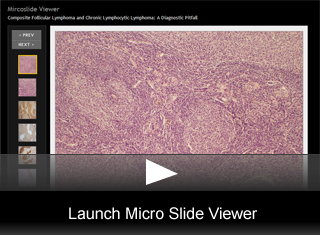
Hematopathology Case 01
78-Year Old Male with Supraclavicular Lymphadenopathy: A Diagnostic Pitfall
by Archana M. Agarwal, MD, Assistant Professor of Pathology, University of Utah School of Medicine
Medical Director, ARUP Laboratories
Nikhil A. Sangle, MD and Barnali Deka, MD.
Editor: Mohamed E. Salama, MD, Associate Professor (Clinical) of Pathology, University of Utah School of Medicine
Medical Director, ARUP Laboratories
A 78- year old male presented with a 2.0cm diameter enlarged left supraclavicular lymph node. The patient had a previous diagnosis of chronic lymphocytic leukemia (CLL). The patient had been recently feeling worse and a left supraclavicular lymph node biopsy was performed to rule out either transformation of the CLL to a large cell lymphoma or some other disease process.
Description:
Pathology Findings:
- Hematoxylin & eosin stained sections of the left supraclavicular lymph node showed a complete effacement of the architecture by atypical follicles and interfollicular expansion.
- The follicles lacked well-defined mantle zones as well as polarity.
- They were variably sized and were composed predominantly of small-cleaved cells with irregular nuclear contours, clumped chromatin and inconspicuous nucleoli.
- Interfollicular areas showed proliferation of predominantly small lymphocytes with oval to round nuclei, and clumped chromatin (fig. 1) (fig. 2).
- The lymphoproliferative process was also seen outside the capsule.
Immunophenotype Findings:
- Immunohistochemical stains for CD3, CD20, CD5, BCL-6, CD10, MIB-1 and BCL-2 were performed with adequately reactive controls.
- CD20 stain highlighted the follicles as well as the interfollicular areas.
- However, the intensity of the CD20 stain was much weaker in the atypical cells of the interfollicular areas (fig. 3).
- The cells of the interfollicular areas also showed co-expression of CD5 (fig. 4).
- BCL-2 stain was strongly positive on the majority of the follicles and weakly positive on the cells of the interfollicular areas (fig. 5).
- CD10 stain highlighted the cells of the follicles (fig. 6).
- Multiple attempts to perform FISH studies to ascertain relationship between these two lymphomas failed due to technical reasons.
- The case was diagnosed as a composite follicular lymphoma and chronic lymphocytic lymphoma.
Final Diagnosis & Discussion
© The copyright for photographs and digital images shown in this case report is owned by ARUP Laboratories, Inc. Unlicensed publication in print, on the internet, or in any other media form of these digital images or photomicrographs for any purpose without written permission is strictly prohibited. Limited use for teaching is permitted. Please contact The Webmaster for licenses and permissions. If you wish to use these images as aid in lectures or scientific slide presentations, each image should accompany the following text: "copyrighted material: www.arup.utah.edu"