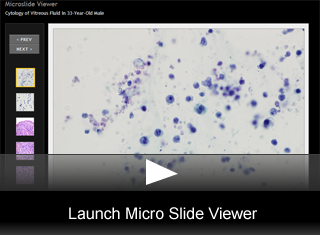
Cytology of Vitreous Fluid in 33-Year-Old Male
by Vasiliki Leventaki, MD, Pathology Resident and KaLynne Harris, MD, Dermatology Resident, University of Utah.
Editor: Brian T. Collins, MD, Professor of Pathology, University of Utah, and Medical Director, Cytopathology, ARUP Laboratories
A 33-year-old male presented at the Dermatology Clinic for diffuse and spreading skin lesions over the past 3 months. Physical examination revealed diffuse pink-to-purple nodules and tumors scattered on his head and neck, trunk and extremities, the largest of which was more than 20 cm in greatest dimension and ulcerated. He also had marked gingival hypertrophy. There was not significant past medical history.
As part of the work up a PET scan was performed that also revealed multiple lung nodules concerning for disseminated disease. A skin biopsy was performed that revealed CD4+ large cell lymphoma. The patient was subsequently admitted to the Hematology/Oncology Clinic and was treated for peripheral T-cell lymphoma, stage IVB. During the course of the chemotherapy treatment, he complained for blurry vision and an ophthalmology evaluation showed vitreitis. A diagnostic vitrectomy was performed and 50ml of clear fluid were submitted to cytology lab for processing and examination. Cultures for fungal and bacterial organisms were also performed on the specimen and were negative.
Cytomorphology Description:
Cytologic Features:
- The cytology laboratory received 50 mL of clear fluid designated as vitreous fluid; one filter and one liquid-based slide were prepared, both stained with Papanicolaou stain.
- Both the filter and the cytospin showed an increased cellularity for vitreous fluid e and atypical lymphoid cells which nuclear membrane irregularities and prominent nucleoli (fig. 2).
- These findings were considered suspicious for involvement by lymphoproliferative disorder.
- Due to the cytologic findings and the pertinent history of peripheral T-cell lymphoma, a portion of the specimen was submitted for flow cytometric studies.
- Immunophenotypic analysis revealed a paucicellular specimen with creased viability and cellular debris that also included a small population of atypical T-cells that appeared to express CD3, weak CD2 and partial CD7, but not CD4, CD8 or CD30.
- These findings were suspicious for T-cell disorder, but not definite due to the decreased viability of the specimen.
Histologic Features:
- At the time of initial presentation two skin punch biopsies were received in the histology lad that were submitted.
- Microscopic examination of the punch skin biopsies revealed a dense, dermal infiltrate of large, atypical lymphocytes with irregular nuclear contours and nuclear hyperchromasia (fig. 3,fig. 4,fig. 5).
- There was also a focal epitheliotropism of similar appearing lymphocytes.
- PAS and gram stains were performed and were negative for organisms (not shown).
- Immunohistochemical stains were performed in one of the skin biopsies.
- The atypical cells were positive for CD3 (a pan-T-cell marker) and CD2, and positive for CD4, but negative for CD5, CD7, CD8, CD30, CD56, and CD20 (a pan-B-cell marker) (fig. 6).
Final Diagnosis & Discussion
© The copyright for photographs and digital images shown in this case report is owned jointly by Brian Collins, MD and ARUP Laboratories, Inc. Unlicensed publication in print, on the internet, or in any other media form of these digital images or photomicrographs for any purpose without written permission is strictly prohibited. Limited use for teaching is permitted. Please contact The Webmaster for licenses and permissions. If you wish to use these images as aid in lectures or scientific slide presentations, each image should accompany the following text: "copyrighted material: www.arup.utah.edu"
 Site Search


