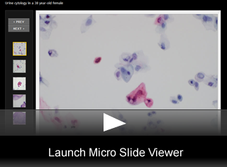
Cytopathology Case 14
Urine cytology in a 38 year-old female
by Leslie E Lopez, MD
Editor: Benjamin L. Witt, MD, Medical Director, Cytopathology, ARUP Laboratories, and Assistant Professor of Pathology, University of Utah.
38 year-old female with a background history of T4 paraplegia since 1993 secondary to a motor vehicle accident, with resultant neurogenic bladder, multiple sacral decubitus ulcers and colostomy. The patient has had chronic urinary incontinence and had an indwelling Foley catheter for a prolonged period. She was evaluated for symptoms related to bladder stones and subsequently underwent cystoscopy with bladder biopsy and suprapubic tube placement. Our department received a bladder washing for cytologic evaluation concurrently with the bladder biopsy specimen.
The cytology images are from the bladder washing. The histology images are from the bladder biopsy.
Cytomorphology Description:
Microscopic Features:
- High power smear showing small single cells with nuclear hyperchromasia and irregular nuclear contours as well as cell clusters composed of larger cells dense, uniform cytoplasm. Some of the nuclei within the larger cells are enlarged, irregularly shaped and dark.(fig. 1)
- A higher power view shows a cell with a large, hyperchromatic nucleus (left of center). Some of the surrounding cells show nuclear karyorrhexis with orangeophilic cytomplasm.(fig. 2).
- A cluster of cells with orangeophilic cytoplasm and nuclei demonstrating hyperchromasia, enlargement, chromatin clumping and nuclear contour irregularities.(fig. 3).
- Abundant squamous metaplasia of the bladder urothelium (upper) with adjacent areas of full thickness dysplasia (lower).(fig. 4).
- High power view of the urothelium showing full thickness dysplasia, with squamous cell features including superficial keratinization and intercellular bridges. These findings match the features noted on cytology.(fig. 5).
Final Diagnosis & Discussion
© The copyright for photographs and digital images shown in this case report is owned jointly by Benjamin L. Witt, MD and ARUP Laboratories, Inc. Unlicensed publication in print, on the internet, or in any other media form of these digital images or photomicrographs for any purpose without written permission is strictly prohibited. Limited use for teaching is permitted. Please contact The Webmaster for licenses and permissions. If you wish to use these images as aid in lectures or scientific slide presentations, each image should accompany the following text: "copyrighted material: www.arup.utah.edu"
 Site Search


