 Site Search


Fine Needle Aspiration of a Parotid Mass on 57-Year-Old Man
by Julie Ann Walby, MD, Cytopathology Fellow, University of Utah
Editor: Brian T. Collins, MD, Professor of Pathology, University of Utah, and Medical Director, Cytopathology, ARUP Laboratories
A previously healthy 57-year-old male presents to Head and Neck Clinic with a left upper neck mass of 3 months duration. Onset was gradual and painless. There was poor dentition with multiple caries and a history of recent tooth extraction in this same general region. There was no response to antibiotics. The patient works as an outdoor construction worker with sun exposure and has a distant smoking history, 30 years ago. A CT study shows the mass to be 2 cm in size and located within the inferior tail of the parotid gland. A fine needle aspiration (FNA) was performed in clinic.
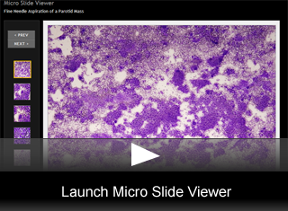
FNA Findings
The modified Wright-Giemsa smears are cellular with a background of hemorrhage and necrotic debris. (fig. 1) The high grade cells are arranged in sheets or small clusters or are present as individual cells. The cells are of variable size and shape including spindled, polygonal, and pleomorphic forms. The nuclei are irregularly round with marked atypia. Many are multinucleated and have bizarre forms. (fig. 2) The cytoplasm is abundant, pale and vacuolated. (fig. 3) Nucleoli are prominent. (fig. 4) The cell block highlights the occasional spindled cell and the abundant eosinophilic cytoplasm. The course irregular chromatin can also be appreciated. (fig. 5)
Final Diagnosis & Discussion
© The copyright for photographs and digital images shown in this case report is owned jointly by Brian Collins, MD and ARUP Laboratories, Inc. Unlicensed publication in print, on the internet, or in any other media form of these digital images or photomicrographs for any purpose without written permission is strictly prohibited. Limited use for teaching is permitted. Please contact The Webmaster for licenses and permissions. If you wish to use these images as aid in lectures or scientific slide presentations, each image should accompany the following text: "copyrighted material: www.arup.utah.edu"