 Site Search


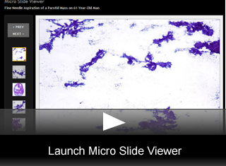
Fine Needle Aspiration of a Parotid Mass on 61-Year-Old Man
by Wells Chandler, MD, Pathology Resident, Department of Pathology, University of Utah.
Editor: Brian T. Collins, MD, Professor of Pathology, University of Utah, and Medical Director, Cytopathology, ARUP Laboratories
A sixty-one-year-old man presented to his primary care physician with an enlarged left parotid gland. The patient was referred to an otolaryngologist, who palpated a firm 2 cm nodule in the region of the left parotid gland. The patient was otherwise asymptomatic. A fine needle aspirate (FNA) was performed and smears and a cell block were prepared.
FNA Findings
Fine needle aspiration shows the following:
- The smears are cellular with many large 3-dimensional aggregates (fig. 1).
- An extensive capillary network, providing a scaffold for the cellular clusters, is prominent at low power (fig. 2).
- A "dirty" cyst fluid background visible at low power (fig. 1).
- Cell debris, degenerating cells and lymphocytes are seen at higher power (fig. 3).
- Crowded cell groups clinging to capillaries and forming papillary structures are frequent; however, salivary ducts are absent (fig. 4).
- Ill formed acinar structures are present and may resemble normal salivary gland.(fig. 5).
- The nuclei are medium sized, uniform and round with evenly dispersed chromatin and sometimes a small nucleolus (fig. 6).
- Some areas on the smear show a mixture of abundant small round lymphocytes and epithelial elements (fig. 7).
- The cell block sections are cellular and show a compressed acinar and papillary architecture. Clear cells are conspicuous in areas of the cell block (fig. 8).
- A mucicarmine stain is negative (fig. 9).
Final Diagnosis & Discussion
© The copyright for photographs and digital images shown in this case report is owned jointly by Brian Collins, MD and ARUP Laboratories, Inc. Unlicensed publication in print, on the internet, or in any other media form of these digital images or photomicrographs for any purpose without written permission is strictly prohibited. Limited use for teaching is permitted. Please contact The Webmaster for licenses and permissions. If you wish to use these images as aid in lectures or scientific slide presentations, each image should accompany the following text: "copyrighted material: www.arup.utah.edu"