Hematopathology Case 02
CD15 positive diffuse large B-cell lymphoma
by Mona Elsakka, MD
PGY-4 Pathology Resident, St. Joseph’s Hospital and Medical Center, Phoenix, AZ
Mohamed Salama, MD
Assistant Medical Director, Hematopathology, ARUP Laboratories, and Associate Professor (Clinical) of Pathology, University of Utah School of Medicine
andRodney R. Miles, MD, PhD
Staff Hematopathologist, ARUP Laboratories, and Assistant Professor of Pathology, University of Utah School of Medicine
Editor: Benjamin L. Witt, MD
Medical Director, Cytopathology, ARUP Laboratories, and Assistant Professor of Pathology, University of Utah School of Medicine
A 49 year old male presented with left supraclavicular lymphadenopathy. An excisional biopsy was performed and the slides and block were referred for consultation.
Description:
Pathology Findings:
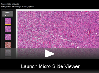
- Hematoxylin & Eosin stained sections of the left supraclavicular lymph node showed complete effacement of the architecture (fig. 1) by sheets of predominantly large, mononuclear cells characterized by irregular nuclear contours, vesicular chromatin and variably prominent nucleoli (fig. 2).
- In addition, there were scattered very large, anaplastic cells that showed occasional bi-nucleation and multi-nucleation with occasional Reed-Sternberg (RS) type cells (fig. 3).
- The involved areas did not show nodular growth pattern or significant sclerosis.
- The cellular background consisted of small lymphocytes and occasional histiocytes without significant numbers of eosinophils or plasma cells.
Immunophenotype Findings:
- Immunohistochemical stains for CD45, CD20, CD3, CD10, BCL6, CD15, CD30, CD4, CD8, ALK, OCT-2, PAX-5, BOB.1 and CD79a with appropriately reactive controls were reviewed.
- The atypical large cell population as well as the anaplastic and RS type cells strongly expressed CD20 (fig. 4), CD79a (fig. 5), PAX-5 (fig. 6), and OCT-2 (fig. 7) with weaker BOB.1 expression(fig. 8).
- The scattered anaplastic and RS-like cells showed weak and variable reactivity with CD45 (fig. 9) as well as variable reactivity with CD30 (fig. 10) and CD15 (fig. 11).
- CD30 was weak to moderate on some of the large cells while many were negative.
- CD15 was strong in some of the large cells while others were negative.
- ALK was negative.
- CD3 highlighted background T lymphocytes, which included a mixture of CD4 and CD8 subsets.
Final Diagnosis & Discussion
© The copyright for photographs and digital images shown in this case report is owned by ARUP Laboratories, Inc. Unlicensed publication in print, on the internet, or in any other media form of these digital images or photomicrographs for any purpose without written permission is strictly prohibited. Limited use for teaching is permitted. Please contact The Webmaster for licenses and permissions. If you wish to use these images as aid in lectures or scientific slide presentations, each image should accompany the following text: "copyrighted material: www.arup.utah.edu"
 Site Search


